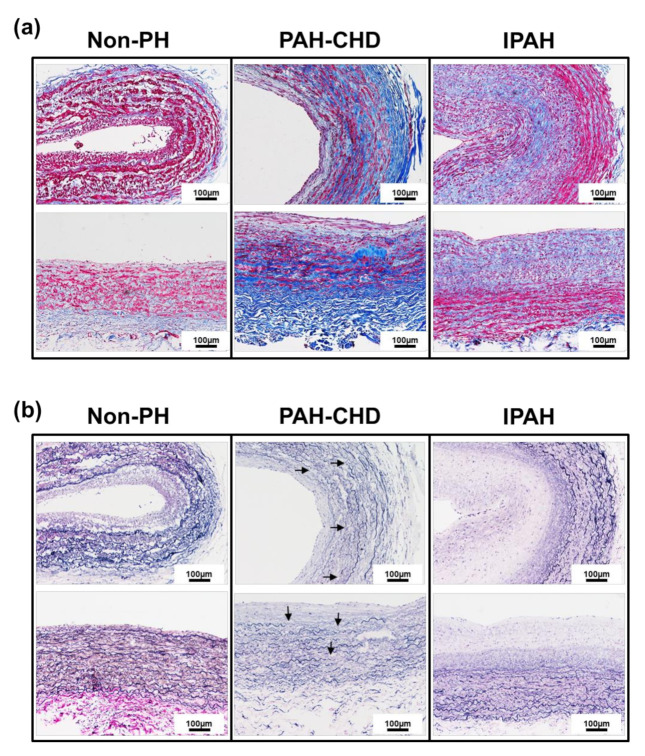
Figure 1.
Collagen and elastin fiber distribution in the PAs of non-PH, PAH-CHD and IPAH patients. (a) Representative the Masson’s trichrome collagen staining in the pulmonary arteries PAs from the non-PH, PAH-CHD and IPAH patientpatients. The collagen content in the PA tissue from a PAH-CHD patient was significantly increased in the medial and adventitial layers compared with that in tissues from IPAH and non-PH patients. (b) Representative modified Verhoeff elastin-stained PAs in these three groups. Internal and external elastic laminae (black staining) are apparent in the non-PAH PA patient, but loss of elastin and fragmentation (arrows) are observed in the PAH-CHD patient (Scale bar: 100 µm).

