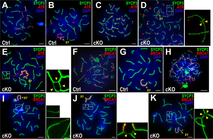
Fig. 6. Impaired ATR and BRCA1 signals in Bruce cKO spermatocytes with chromosomal translocations.
Immunofluorescence (IF) staining of SYCP3 (green) and ATR (red) in chromosome spreads of Bruce Ctrl and cKO spermatocytes in the zygotene (a, c) and pachytene (b, d, e) stages. We observed the normal formation of ATR foci in Bruce cKO zygotene spermatocytes (c), the partial loss of ATR signals at unsynapsed chromatin within the radials of Bruce cKO mid pachytene spermatocytes (d), and total loss of ATR signals at unsynapsed segments in Bruce cKO late pachytene spermatocytes (e). The chromosomal radials within the dashed rectangles are magnified to the right (arrowheads, unsynapsed chromosomal segments). Note that ATR signals on the sex chromosomes (yellow arrows) are also reduced (also see Fig. S4A–C). Representative images from two independent Bruce Ctrl-cKO littermate pairs (cKO spermatocytes bearing radials, n = 21). Bars, 5 µm. IF staining of SYCP3 (green) and a marker of chromosomal synapsis, BRCA1 (red), in chromosome spreads of Bruce Ctrl and cKO spermatocytes in the zygotene (f, h) and mid pachytene (g, i) stages. BRCA1 accumulation patterns appear normal in Bruce cKO zygotene spermatocyte (h), while are largely lost in Bruce cKO pachytene spermatocytes bearing chromosomal fragments (i), which are bounded by dashed rectangles and magnified to the right. Note that BRCA1 signals are largely or entirely absent from the cKO mid pachytene chromosomal fragments. Representative images from two independent Bruce Ctrl-cKO littermate pairs. Bars, 5 µm. (j, k) IF staining of SYCP3 (green) and BRCA1 (red) in Bruce cKO spermatocytes bearing chromosomal translocations in the mid pachytene (j) and late pachytene (k) stages. Chromosomal radials in the dashed rectangles are magnified to the right. Note that, within the radials, BRCA1 signals are partially lost from unsynapsed regions in the mid pachytene stage and entirely lost from such regions in the late pachytene stage (arrowheads), while BRCA1 signal appears normal on the sex chromosomes in both stages (yellow arrows). Representative images from three independent Bruce Ctrl-cKO littermate pairs (cKO spermatocytes bearing radials, n = 16). Bars, 5 µm.

