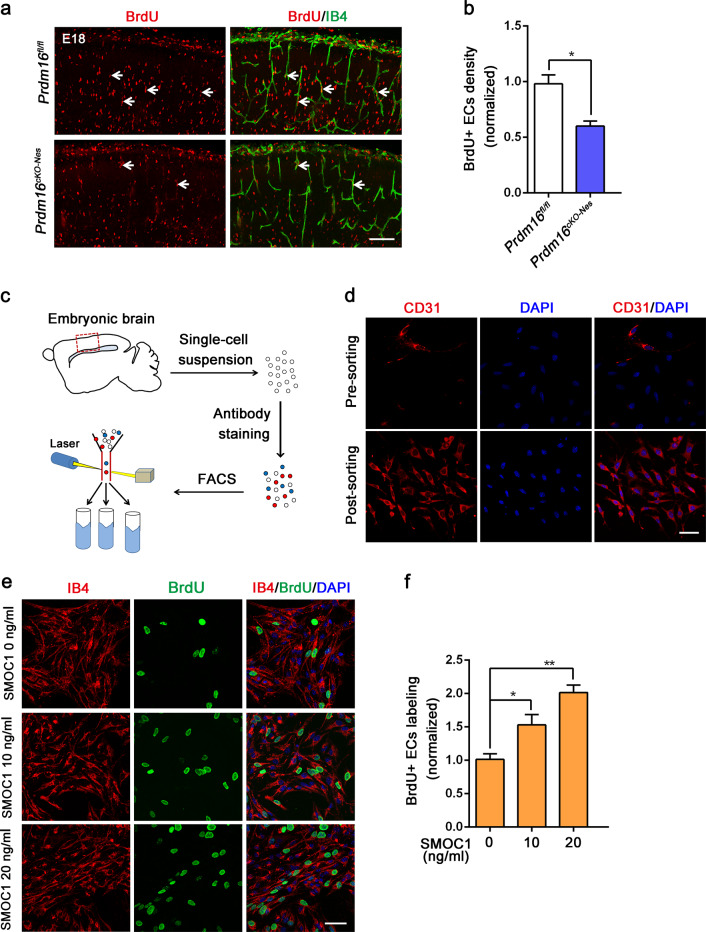
Fig. 5. Prdm16 deletion disrupts vascular proliferation, and SMOC1 promotes ECs proliferation.
a Representative confocal images of cortical ECs proliferation at E18.5. A reduced number of BrdU-labeled ECs was observed in the Prdm16cKO-Nes cortical plate. Scale bar, 100 μm. b Quantification of the decrease in the number of BrdU+ ECs when compared with that in the Prdm16fl/fl mouse cortex. The data are represented as the means ± SEM (n = 3 animals; unpaired Student’s t test; *P < 0.05). c Schematic overview of the strategy used to sort ECs in cell suspensions with a FACSCalibur cytometer. d Representative confocal images of cell suspensions costained with CD31 and DAPI presorting and postsorting. e Immunofluorescence staining with IB4/BrdU in ECs showed increased endothelial cell proliferation upon the addition of SMOC1 (0, 10, 20 ng/ml) to the culture medium. Scale bar, 50 μm. f Quantification of the increased proliferation of BrdU-positive ECs upon SMOC1 (0, 10, 20 ng/ml) application. The data are represented as the means ± SEM (n = 3; unpaired Student’s t test; *P < 0.05; **P < 0.01).

