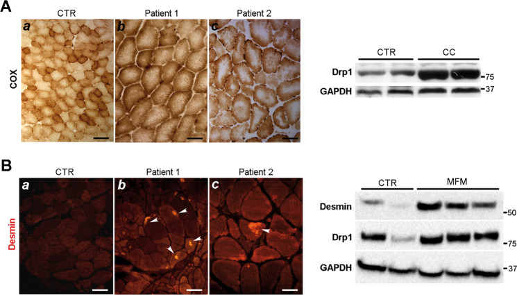
Fig. 8. Central core and myofibrillar myopathy show high Drp1 expression.
a Central Core (CC) myopathy left panel: COX staining in two human muscle samples showing central area without or reduced COX activity (b–c). (a) COX activity in normal human muscle samples. Scale Bar (a: 50 µm, b–c: 25 µm). Right panel: Drp1 immunoblotting in muscle homogenates from controls and CC human biopsies. b Myofibrillar myopathy (MFM) left panel: desmin immunostaining showing desmin accumulation in two human muscle samples (white arrows) (b–c). (a) Desmin immunostaining in normal human muscle sample. Scale Bar (a: 40 µm, b–c: 25 µm). Right panel: Drp1 immunoblotting in muscle homogenates from controls and MFM human biopsies.

