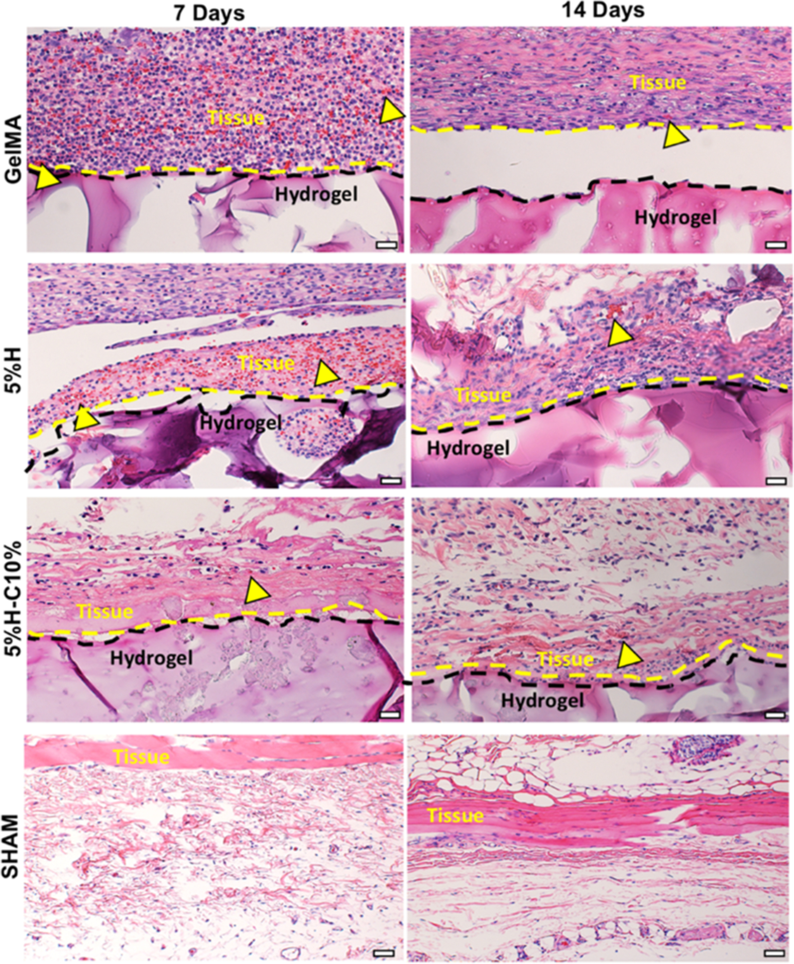
Figure 10.

Histological analysis of the implanted hydrogels. Representative H&E staining images of GelMA, 5%H, 5%H-C10%, and SHAM explants (hydrogels with the surrounding tissue) after 7 and 14 days in vivo (scale bar = 200 μm). Yellow arrowheads indicate the presence of numerous blood vessels containing murine erythrocytes.
