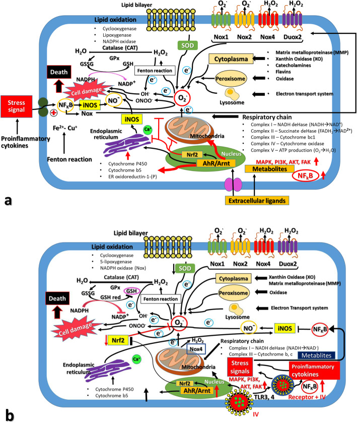
Fig. 1.
Schematic representation of ROS/RNS production and Redox control in response to stresses inducing reagents and influenza virus (IV) infection. Subcellular organelles and cellular components were affected by the oxidation and antioxidation responses in the case of stress inducers like phase I reagents and environmental hormones (Panel a) and infection of IVs (Panel b). The source of cellular ROS/RNS production mainly occurred in mitochondria, endoplasmic reticulum, lipid bilayer, organelle membranes, cellular lipids, DNA in the nuclei, and cellular oxidation enzymes and antioxidation enzymes such as superoxide dismutases (SODs), catalases (CATs), peroxidases (Prdxs), glutathione peroxidase (GPx) and glutaredoxins (GRs) and so on. In addition, the cellular signaling pathways such as NF-kB, MAPK, PI3K, AKT and iNOS signaling are also shown. After IVs infection, NOX produced superoxide (O2-) and dysfunction on the mitochondrial proteins. The defective mitochondrial proteins resulted in the leakage of electrons and superoxides from the mitochondria, as well as initiating cell death pathways by cytochrome c or permeability transition pore (PTP). The NF-kB produced many cytokines as well as inducible nitric oxide (NO) synthase (iNOS). This iNOS then produced nitric oxides (NO). The NO and O2- reacted together to produce peroxynitrite (ONOO) which is a highly reactive compound to generate the protein nitration and damage of macromolecules. and viral mutations. Higher generation of O2- resulted in the production of H2O2 by the catalytic activity of SOD. Uncontrolled production of H2O2 produced hydroxyl radicals (OH-) via reaction with metal cations, and these H2O2 and OH- caused irreversible damages to cellular proteins, lipids, nucleic acids and so on. This Figure is a modified version of the ones published by Di Meo et al. [16] and Kohmich et al. [46].

