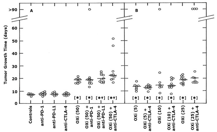
Figure 2.
Effect of OXi4503 (OXi) and monoclonal antibody inhibitors (10 mg/kg) of PD-1, PD-L1, and CTLA-4 on the growth of a C3H mammary carcinoma implanted in CDF1 mice. These mice were i.p. injected with different drugs, with the treatments starting when tumors had reached a volume of 200 mm3 (day 0). The actual treatment days were 0, 3, 7 and 10 (OXi) or 1, 4, 8 and 11 (checkpoint inhibitors). Results are individual values with the line indicating the median for each group and show the tumor growth time (time for tumors to reach 5 times the starting treatment volume). Values at >90 days indicate those tumors that were controlled so no actual tumor growth time value was possible. For (A), the results are for control animals, mice treated with each checkpoint inhibitor (10 mg/kg) alone, a high OXi dose (50 mg/kg) alone, or OXi and each inhibitor combined. (B) shows results using lower OXi doses (5, 10, or 25 mg/kg) with/without anti-CTLA-4. The different OXi doses are shown in the parentheses on the x-axis. Statistical comparisons of the data in both figures were made using a Wilcoxon-Mann-Whitney test and show those groups that were significantly different (p < 0.05) from controls [*] or each respective OXi dose alone [†].

