Fig. 14.2.

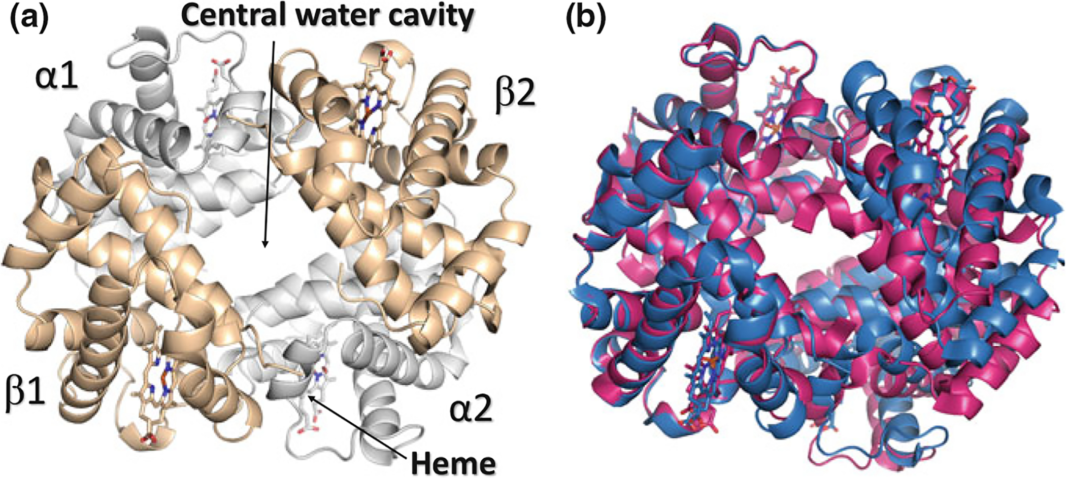
Crystal structure of hemoglobin. a Overall quaternary structure of Hb with the two α chains and β chains colored grey and tan, respectively. b Structure of oxygenated (R state) Hb (magenta) superimposed on the structure of deoxygenated (T state) Hb (blue). Note the larger central water cavity in the T structure
