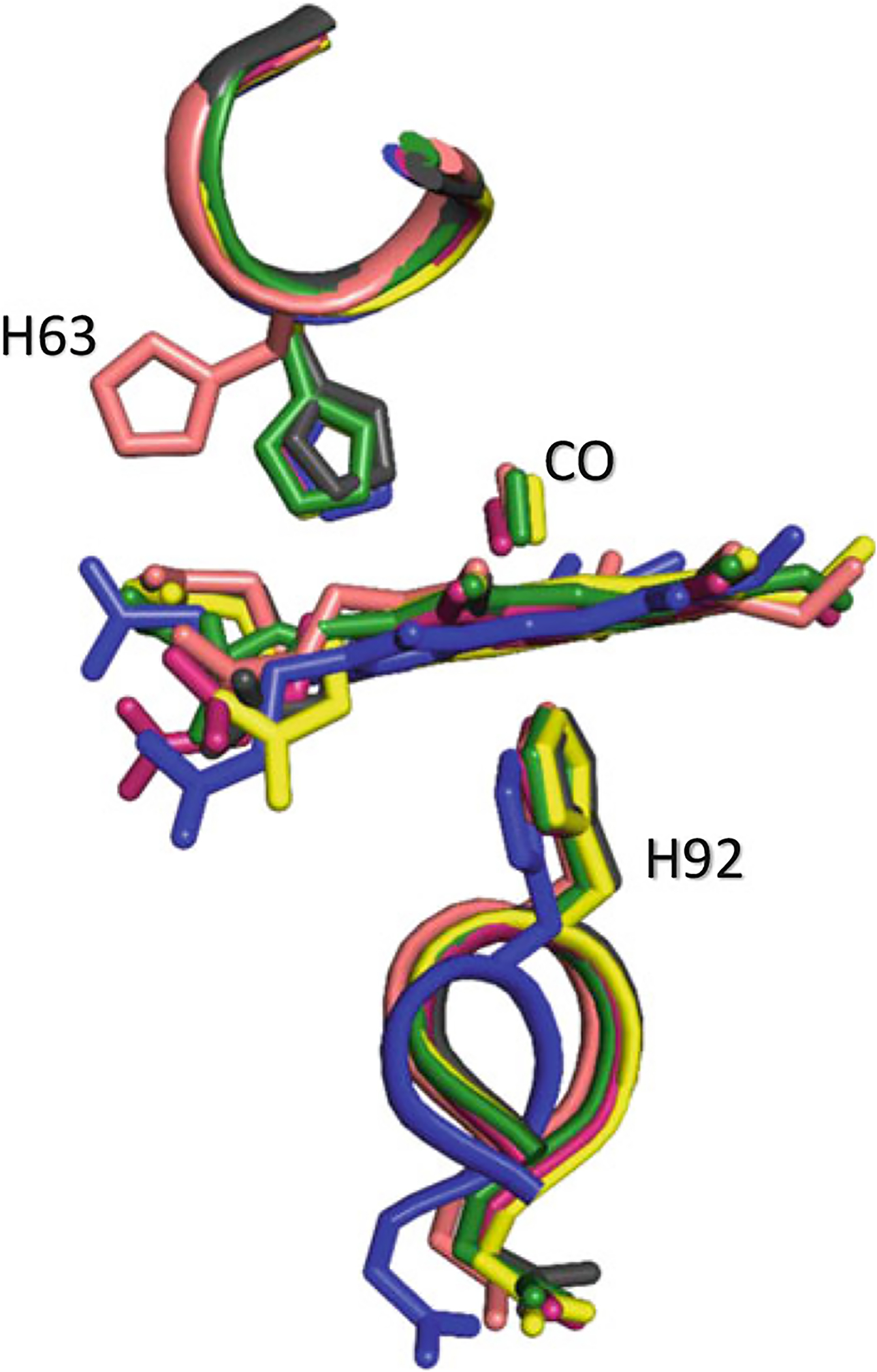
Fig. 14.4.

Superposed β heme structures of R (magenta), R3 (yellow), RR2 (green), R2 (black), and RR3 (salmon) showing the positions of βHis63. Note the rotation of βHis63 out of the distal pocket in the RR3 structure, creating a ligand channel to the bulk solvent, while in R, RR2, and R2 structures, βHis63 is still located in the pocket making hydrogen-bond interaction with the ligand. The R3 structure shows a partially opened ligand channel
