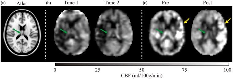
Figure 4.
Longitudinal cerebral blood flow changes in two representative patients. (a) A T1-weighted atlas at the approximate location of the shown blood flow maps. (b) A patient with moyamoya (age = 58 years; sex = female) scanned at an interval of 72 days with no interim surgery. The images suggest subtle reductions in cortical perfusion and slight increases in choroid plexus perfusion (green arrow). (c) A patient with moyamoya with interval surgery (age = 31 years; sex = female) scanned 72 days after left-sided encephaloduroarteriomyosynangiosis. Increases in cortical perfusion are observed near the revascularization site (yellow arrow), whereas bilateral decreases in choroid plexus perfusion are observed.

