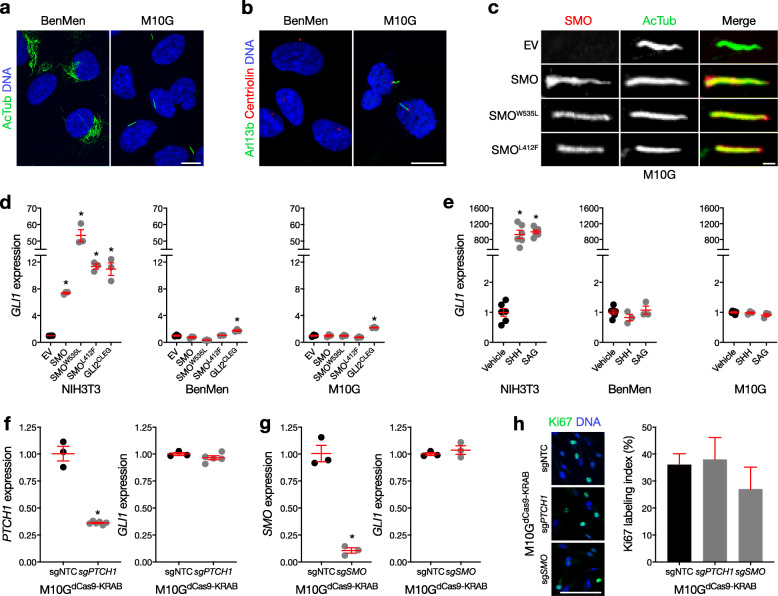
Fig. 2.
Primary meningioma cells accumulate Smoothened in cilia, but do not transduce ciliary Hedgehog signals. a, b Confocal immunofluorescence microscopy for the ciliary markers acetylated tubulin (AcTub) or Arl13b, the ciliary base/centriole marker Centriolin, and DNA (Hoechst) reveals that M10G meningioma cells express primary cilia, but BenMen meningioma cells do not, instead expressing AcTub in a perinuclear pattern. c Confocal immunofluorescence for SMO 72 h after transfection of SMO, oncogenic point substitutions of SMO (SMOW535L or SMOL412F), or empty vector (EV) control, shows that exogenous SMO accumulates in the cilia of M10G meningioma cells. d qRT-PCR assessment of GLI1 expression 72 h after transfection of SMO, SMOW535L, SMOL412F, constitutively active GLI2 (GLI2CLEG), or EV control, shows that over-expression of SMO activates the Hedgehog transcriptional program in NIH/3T3 cells, but not in BenMen or M10G meningioma cells. In contrast, over-expression of GLI2CLEG mildly induces GLI1 expression in meningioma cells (*p < 0.01 Student’s t test). e qRT-PCR assessment of GLI1 expression 24 h after treatment with recombinant Sonic Hedgehog (SHH, 1 μg/ml), SMO agonist (SAG, 100 nM), or vehicle control, demonstrates that pharmacologic stimulation of the Hedgehog pathway activates the Hedgehog transcriptional program in NIH/3T3 cells, but not in BenMen or M10G meningioma cells (*p < 0.0001 Student’s t test). f qRT-PCR assessment of GLI1 expression shows that CRISPRi suppression of PTCH1 (sgPTCH1) compared to transduction of non-targeted sgRNAs (sgNTC) fails to activate the Hedgehog transcriptional program in M10G meningioma cells (*p < 0.0001 Student’s t test). g qRT-PCR assessment of GLI1 expression shows that CRISPRi suppression of SMO (sgSMO) compared to transduction of sgNTC fails to suppress the Hedgehog transcriptional program in M10G meningioma cells (p < 0.0001 Student’s t test). h Quantitative confocal immunofluorescence microscopy for the proliferation marker Ki67 demonstrates that CRISPRi suppression of PTCH1 or SMO compared to sgNTC fails to alter M10G meningioma cell proliferation. qRT-PCR data are normalized to GAPDH expression and EV, vehicle, or sgNTC controls as indicated. All experiments were performed at least 3 times with at least 3 technical replicates

