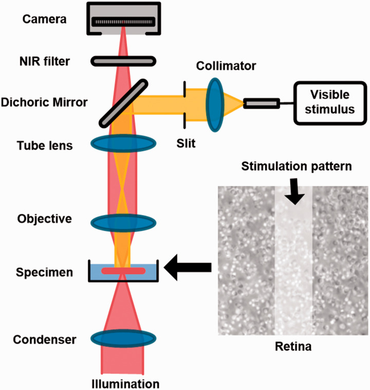
Figure 1.
Schematic diagram of the experiment setup. A custom modified microscope, with a 60× objective and a CMOS camera (16 bits depth and 100 frames/s), was used for retinal imaging. During the experiment, the photoreceptor outer segment (OS) tips were facing upward and the retina was perfused with oxygenated physiological solution (pH 7.4 and 35–37°C). A rectangular stimulus pattern, generated by a slit and a LED at visible wavelength, was projected to the retina for localized stimulus. The stimulus duration was 500 ms for all the experiments. (A color version of this figure is available in the online journal.)

