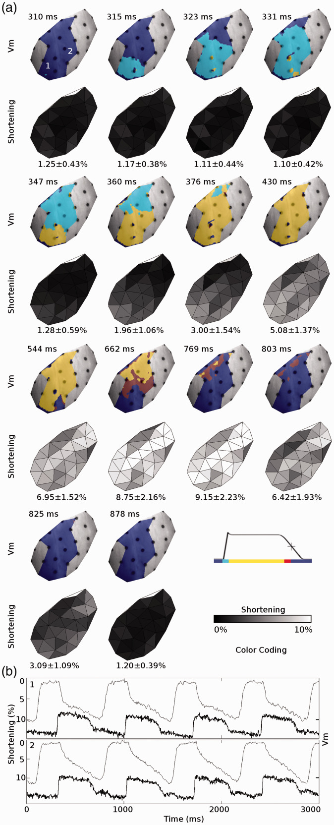
Figure 2.
Electrical and mechanical functions mapped simultaneously in a beating ex-vivo pig heart using the method of Zhang et al.15 (a: upper rows) Propagation of membrane depolarization and repolarization for a single apically paced beat. (a: lower rows) The resulting contraction. The mapped region includes the anterior left ventricle. The left anterior descending coronary artery is along the top-left edge of each image and the apex is at the bottom. (b) Membrane potential (Vm, bold line) and mechanical shortening (fine line) recorded from the two sites indicated in (a). Shortening is defined as the most negative eigenvalue of the stretch tensor. Reproduced from Zhang et al.15 with permission.

