Figure 1.
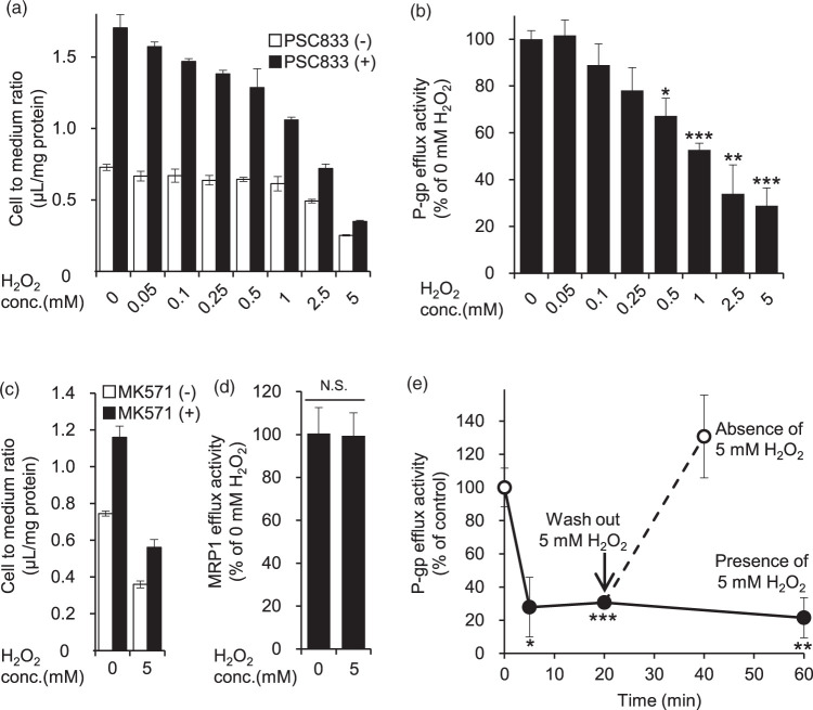
Effect of H2O2 on P-gp- and MRP1-mediated [3H]vinblastine efflux activity in hCMEC/D3 cells. (a, c) hCMEC/D3 cells were pre-incubated in extracellular fluid (ECF) buffer containing DMSO, 10 µM PSC833 (P-gp inhibitor) or 50 µM MK571 (MRP1 inhibitor) for 30 min at 37℃, and then the cellular uptake of the P-gp substrate vinblastine was measured for 20 min in the presence of H2O2 (0.05 mM to 5 mM) or ECF buffer (0 mM H2O2). To effectively inhibit P-gp or MRP1 efflux activity, PSC833 or MK571 was present during both the 30 min pre-incubation and the 20-min uptake period. The cellular uptake was expressed as the cell-to-medium ratio, as described in Materials and methods. Each column represents mean ± SD (n = 3). (b, d) Based on the data of Figure 1(a) and (c), P-gp or MRP1 efflux transport activity was calculated from the cell-to-medium ratio in the absence and presence of PSC833 or MK571 according to Materials and methods. (e) Cellular uptake of vinblastine was measured at 5 min, 20 min and 60 min after addition of 5 mM H2O2 (closed circles). To determine the effect of 5, 20 or 60 min treatment with H2O2 on the P-gp efflux transport activity, hCMEC/D3 cells were pre-incubated in ECF buffer containing DMSO or 10 µM PSC833 for 30 min at 37℃, and then the cellular uptake of vinblastine was measured for 5, 20 or 60 min with or without 5 mM H2O2. To examine reversibility, the medium was replaced with fresh medium after pre-incubation with 5 mM H2O2 for 20 min (open circles), and the cellular uptake of vinblastine was measured for 20 min in the absence of 5 mM H2O2. Both pre-incubation and vinblastine uptake were conducted in the presence of DMSO or PSC833. Each value represents the mean ± SD (n = 3). Statistical significance was determined by one-way ANOVA followed by post hoc Bonferroni-corrected Student's t-test. *p < 0.05, **p < 0.01, ***p < 0.005.

