
Figure 2.
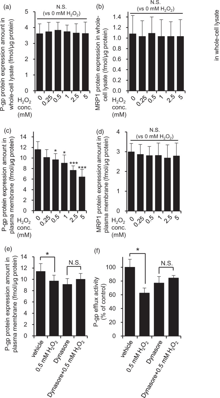
H2O2 facilitated dynamin-dependent P-gp internalization in hCMEC/D3 cells. The effect of H2O2 on P-gp and MRP1 protein expression amounts in whole-cell lysate (a, b) and plasma membrane fraction (c, d) was examined. hCMEC/D3 cells were exposed to 0.25 mM to 5 mM H2O2 for 20 min in ECF buffer. Whole-cell lysate and plasma membrane fraction of hCMEC/D3 cells were digested with lysyl endopeptidase and trypsin. The digests were subjected to LC-MS/MS together with internal standard peptides. Each value represents the mean ± SD of 8–16 SRM/MRM transitions in three to four independent samples. One-way ANOVA with Bonferroni-corrected Student's t-test was performed to determine the statistical significance of differences between protein expression levels under control and H2O2-treated conditions. (e) Dynasore attenuated the decrease of P-gp protein expression amount in the plasma membrane fraction caused by 0.5 mM H2O2 exposure. hCMEC/D3 cells were pre-incubated with dynasore (80 µM) or DMSO (vehicle) for 30 min, and then co-incubated with 0.5 mM H2O2 and dyansore or DMSO in ECF buffer for 20 min at 37℃. P-gp protein expression amounts in plasma membrane fraction were determined by LC-MS/MS analysis as described above. Each value represents the mean ± SD of 9-12 SRM/MRM transitions in three independent samples. (f) Dynasore attenuated the decrease of P-gp efflux transport activity caused by 0.5 mM H2O2 treatment. hCMEC/D3 cells were pre-incubated with dynasore (80 µM) for 30 min, and then the cellular uptake of vinblastine was measured with or without 0.5 mM H2O2, PSC833 and dynasore for 20 min. Each value represents the mean ± SD (n = 3). *p < 0.05, ***p < 0.005 (Student’s t-test).