Figure 1.
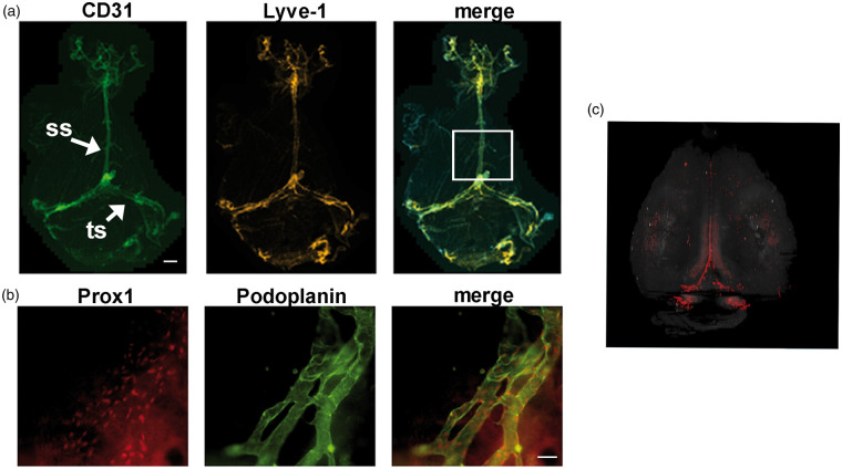
Whole mount imaging of meningeal lymphatic vessels in mice. (a) Meningeal LVs were characterized by whole-mount immunofluorescence staining for CD31 (green) and Lyve-1 (red). Low magnification view of meningeal LVs. LVs run alongside the sagittal sinus (ss) and transverse sinus (ts). The boxed regions in panel A outline the areas that we analyzed in our stroke experiments to acquire lymphatic vessel index (LVI) at the ss. (b) Representative images showing Prox1 (red) and podoplanin (green) expression by meningeal LVs around the transverse sinus (TS). (c) A 3D rendering of meningeal LVs (red) in a Prox1-tdTomato transgenic mouse was generated using serial two-photon tomography. The signal from the hippocampus was digitally removed. (a), scale bar is 1000 µm; (b), scale bar is 20 µm.

