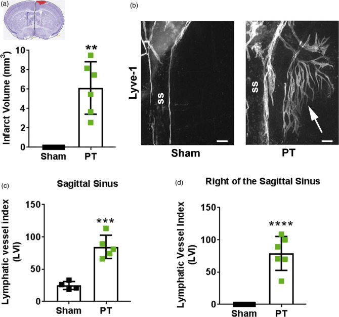
Figure 3.
Photothrombosis induces meningeal lymphangiogenesis. (a) Infarct volumes in mice that received photothrombosis (PT; green squares; n = 6) compared to uninjured sham mice (black squares; n = 4). Inset shows representative cresyl violet-stained cortical section with infarct highlighted in red. (b) Representative image of lymphangiogenesis into the right sagittal sinus (ss) after PT (white arrow). (c) Quantification of meningeal lymphatic vessels at the sagittal sinus and (d) to the right of the sagittal sinus two weeks after PT stroke (n = 5-6) or after sham surgery (n = 4). All data presented as mean ± SD and were analyzed using a Mann–Whitney test. **p < 0.01, ***p < 0.001, ****p < 0.0001. Scale bar = 200 µm.

