Figure 5.
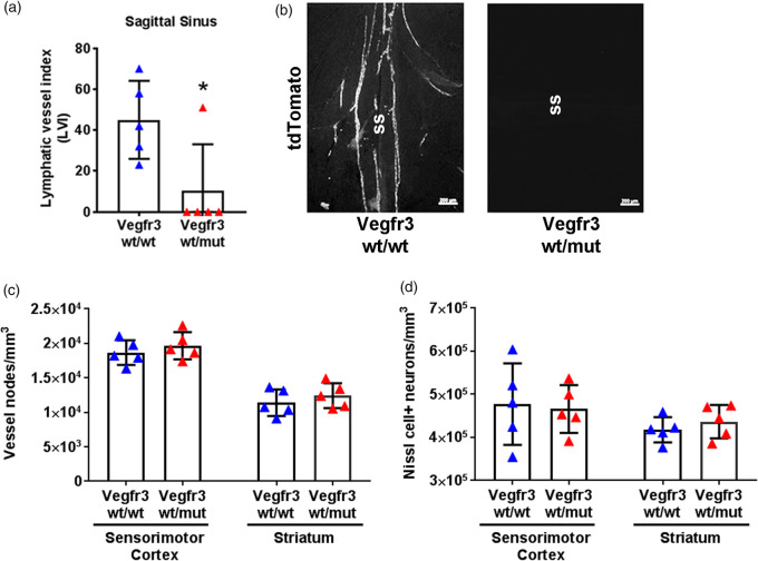
Meningeal hypoplasia does not affect vascular or neuronal development. (a) Quantification of LVs at the sagittal sinus in naïve Vegfr3wt/wt (blue triangles) and Vegfr3wt/mut mice (red triangles) demonstrate meningeal lymphatic hypoplasia. (b) Representative images of tdTomato+vessels at the sagittal sinus (ss). (c) Stereological quantification of microvessel density in the sensorimotor cortex (p = 0.55) and striatum (p = 0.42) of Vegfr3wt/wt and Vegfr3wt/mut mice and (d) stereological quantification of neuronal counts in the sensorimotor cortex (p>0.99) and striatum (p = 0.55) (n = 5 per group). All data presented as mean ± SD and were analyzed using a Mann–Whitney test or two-way ANOVA. *p < 0.05. Scale bar = 200 µm.

