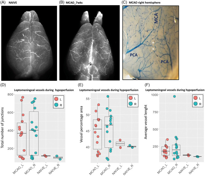
Figure 6.
(A–F) Leptomeningeal vessels in MCAO mice seven weeks post-surgery. Leptomeningeal vessels in naïve mouse (A) and MCAO (B). (C) Anastomoses between terminal branches of PCA and MCA in the right cortex of MCAO mice. MCAO mice are characterized by a symmetric network of leptomeningeal arterioles with increased anastomoses (D), density (E) and moderately increased vessel length (F), both in cortex ipsilateral and contralateral to MCAO. MCA: middle cerebral artery; PCA: posterior cerebral artery; L: left; R; right; wks: weeks.

