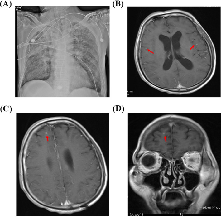
Figure 1.

Chest X‐ray and cranial magnetic resonance imaging (MRI) of the patient with tuberculous meningitis. A, Chest X‐ray showed multiple patches and nodules with observable cavity in his lungs. B‐D, T1‐weighted image showed enhanced nodules in gray matter and white matter border at the right frontal region, as well as lateral meningeal enhancement at the left temporal lobe
