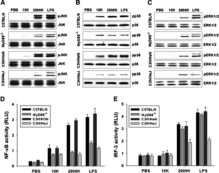
Fig. 3.
Intracellular signaling induced by γ-PGA was MyD88 and TLR4-dependent. Peritoneal macrophages from C57BL/6, MyD88−/−, C3H/HeN, or C3H/HeJ mice were treated with 100 ng/ml LPS and 1 mg/ml γ-PGA for 30 min. The expression level of phosphorylated JNK (pJNK) (a), p38 kinase (pp38) (b), and ERK1/2 (pERK1/2) (c) were determined by Western blot analysis. Peritoneal macrophages from C57BL/6, MyD88−/−, C3H/HeN, or C3H/HeJ mice were transiently transfected with pNF-κB-Luc and IRF-3-Luc reporter gene plasmids and then cultured in complete medium for 24 h. At 24 h after transfection, cells were treated with 100 ng/ml LPS for 1 h and 1 mg/ml γ-PGA for 2 h and then NF-κB activity (d), IRF-3 activity (e) were determined by luciferase reporter assay. The data presented in this figure are representative of triplicate experiments. All data are representative of at least three experiments

