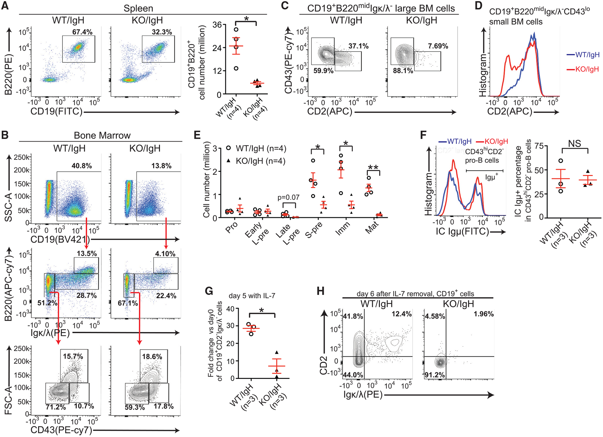
Figure 4. Expression of the Pre-arranged IgH Does Not Rescue B Cell Development Defects in Mettl14 KO Mice.

(A) Flow cytometry plots and quantification of splenic B cells of indicated mice. (B) Flow cytometry plots of B lineage cells in the bone marrow of indicated mice. (C) Expression of indicated markers in the large pre-B cells of indicated mice. (D) Expression of CD2−in the small CD43lo population of indicated mice. (B–D) Data are representative of 4 mice per group. (E) Quantification of indicated B-lineage cells (Figure 1C) in indicated mice. The number of each cell population from 2 femurs per mouse was calculated. (F) Flow cytometry plots and quantification of intracellular Igμ+ cells in indicated cells from indicated mice. (G) The fold changes of CD19+Igκ/λ− CD2−cell numbers were compared between indicated groups after 5-day IL-7-induced expansion. (H) Flow cytometry plots of Igκ/λ−/CD2−expression in CD19+ cells 6 days after IL-7 removal. Data are representative of 3 mice per group. SEM or ± SEM is shown; NS, not significant; *, p < 0.05; **, p < 0.01 and ***, p < 0.001.
