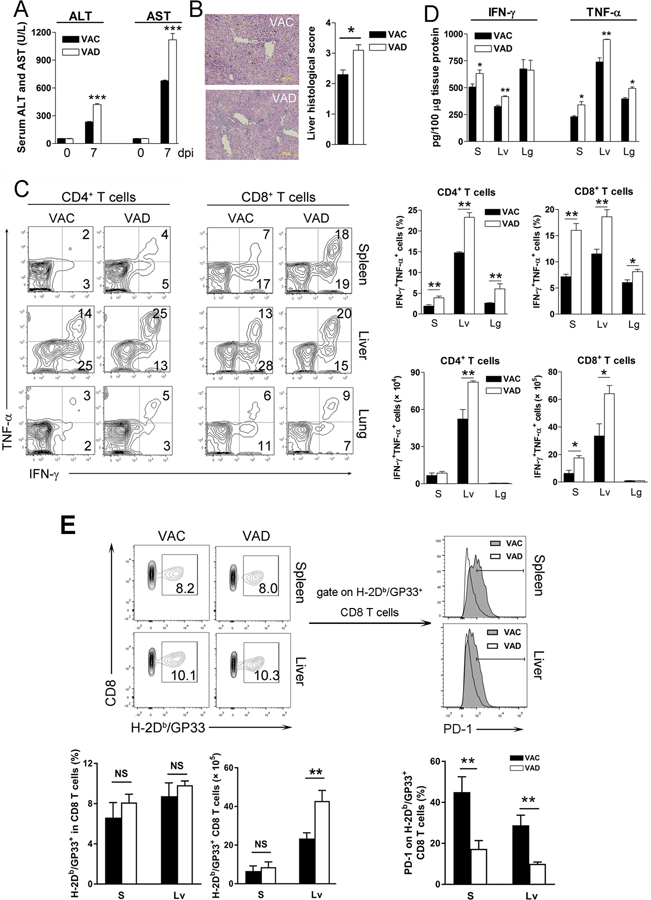
Figure 2. VAD mice exhibited overzealous T cell responses and severe immunopathogenesis at the acute stage of viral infection.
VAC and VAD mice were infected with LCMV Cl13 (2×10⁶ FFU) and sacrificed at 7 dpi. A) Serum ALT and AST. B) Liver histological scores. C) Lymphocytes were isolated from the spleen (S), liver (Lv) and lung (Lg), followed by stimulation with GP33 and GP61 peptides in the presence of BFA for 5 h. Intracellular IFN-γ and TNF-α were analyzed using flow cytometry. D) Liver IFN-γ and TNF-α levels were measured by ELISA kits. E) Virus-specific CD8+ T cells were detected by H-2Db/GP33 MHC tetramer. The PD-1 expression on GP33-tetramer+ CD8+ T cells was measured. The data are shown as mean ± SEM of three to six mice per group from a single representative experiment. The experiment was repeated three times independently. A two-tailed Student’s t-test was used to compare the two groups. A Mann-Whitney test was used to compare the histological scores. * P<0.05, ** P<0.01, *** P<0.001, NS, no significance.

