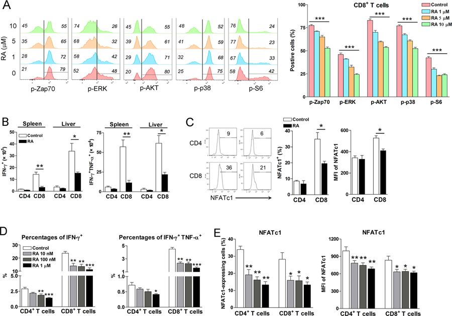
Figure 6. RA inhibited T cell receptor signaling and NFATc1 expression in T cells.
A) Naïve splenocytes were isolated and cultured with RA in vitro for 24 h, followed by the stimulation with anti-CD3 plus anti-CD28 using an antibody- cross-linking method. Cells were fixed immediately by BD Phosflow Lyse/Fix Buffer at 37 °C for 12 mins and permeabilized by BD Phosflow Perm Buffer III on ice for 30 mins. Cells were then incubated with surface antibodies and phosphorylated antibodies for 1 h, followed by flow cytometry analysis. Each group was in triplicates. B) LCMV-infected mice were treated with RA (200 μg/day) at 1, 3, and 5 dpi. The numbers of cytokine-producing T cells and C) the percentages and mean fluorescence intensity (MFI) of NFATc1 were examined at 7 dpi. D-E) Splenocytes of naïve mice were cultured in vitro by anti-CD3/CD28 antibody stimulation in the presence of various concentrations of RA. After 3-day culture, cytokine levels and NFATc1 expression were analyzed by flow cytometry. Each group was in triplicates and the RA treated groups were compared to the control group. All experiments were repeated two to three times independently. A two-tailed Student’s t-test was used to compare the two groups. One-way ANOVA was used to compare more than two groups. * P<0.05, ** P<0.01, *** P<0.001.

