FIG. 2.

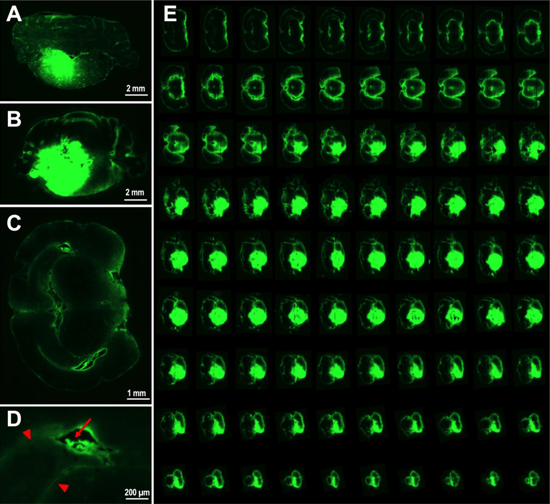
Representative fluorescence (green areas) images of brainstem distribution using 10-kDa FITC-dextran beads after a single 90-μl infusion by CED (A) and 24-hour infusion by osmotic pump delivery (B). Anatomical exploration following CED in the brainstem demonstrated fluorescent parenchymal regions and blood vessels distal from the site of cannulation (C) with distribution along blood vessels (red arrow) and pial interfaces (red arrowheads) observed at higher magnification (D). A summative collage of 7-mm slices following CED with no evidence of reflux (E).
