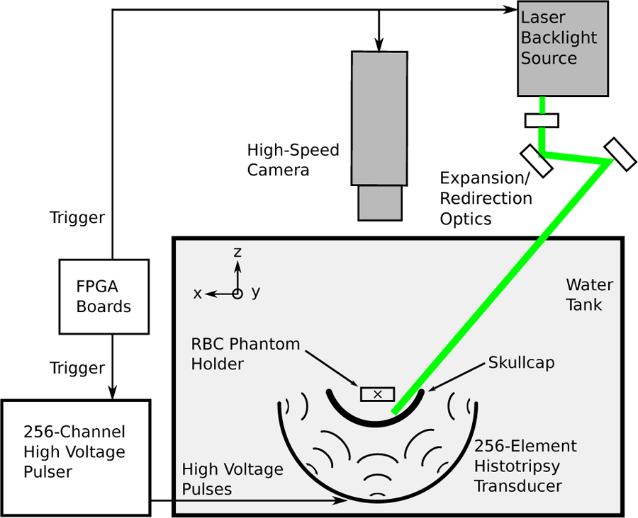
Fig. 3.

Schematic of the experimental setup used for imaging experiments studying single lesion generation through the skullcap. A computer was used to send a trigger signal to the camera and laser for imaging purposes and to the FPGA boards to fire the high voltage pulser responsible for generating the ultrasound pulses used for treatment.
