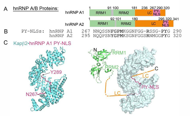
Figure 2. The hnRNP A/B family of proteins are nuclear import cargos of Kapβ2. A, B.
Domain organization (A) and PY-NLS sequences (B) of hnRNP A1 and hnRNP A2. The Kapβ2 binding epitopes of the PY-NLSs are underlined. C. Structure of Kapβ2 (cyan) bound to the PY-NLS of hnRNP A1 (magenta); 2H4M. Kapβ2 is shown with a cartoon representation on the left, and with a surface representation on the right. A schematic of other hnRNP A1 domains are shown in the right panel. Structures of the folded tandem RRM1-RRM2 (2LYV) domains are shown.

