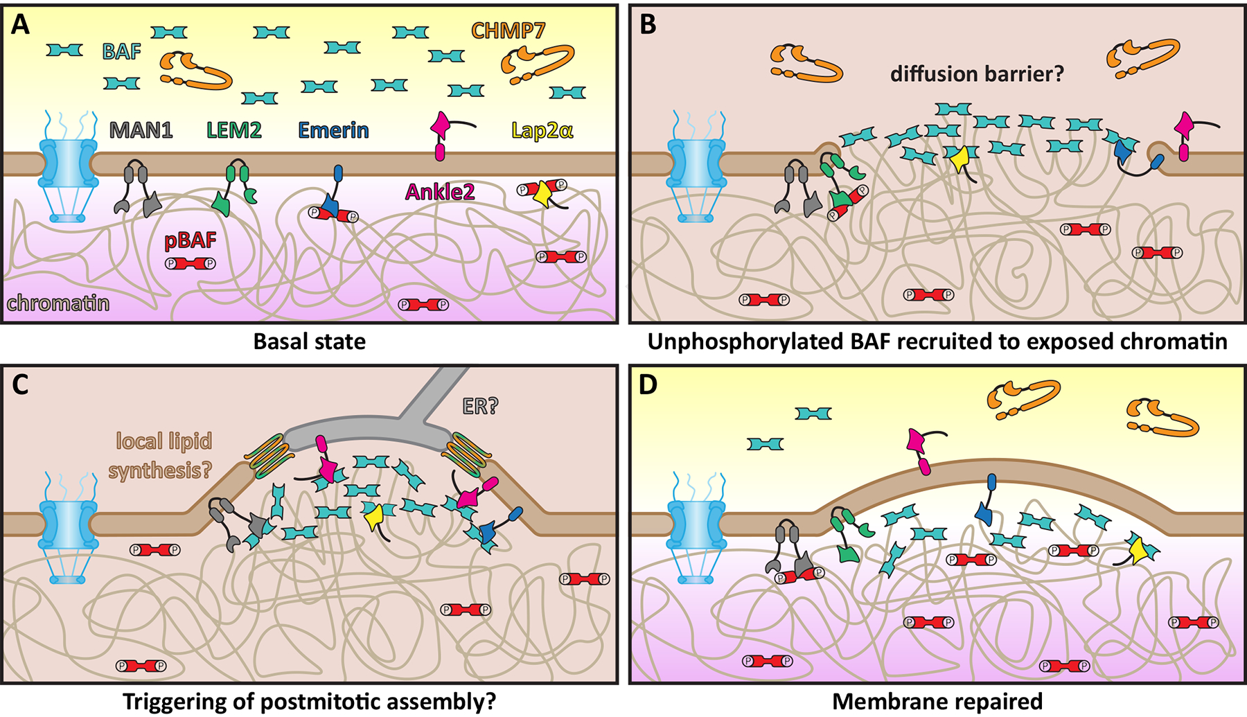
Figure 2. Model for repair of large nuclear envelope ruptures.

A: Schematic of basal/unperturbed state of mammalian nuclear envelope, which is surveilled by CHMP7 (orange) and unphosphorylated BAF (teal). Key proteins involved with surveillance and repair including integral INM LEM proteins MAN1 (grey), LEM2 (green), Emerin (blue), Ankle2 (pink), and Lap2α (yellow) are also shown. Phosphorylated BAF (pBAF, red) is specifically found in the nucleus, bound to LEM domain proteins and chromatin (beige). B: Following nuclear envelope rupture, cytosolic, unphosphorylated BAF binds to exposed chromatin, possibly forming a diffusion barrier. C: LEM domain proteins are recruited to sites of rupture by BAF coating the exposed chromatin. Membrane for repair may be donated from the ER and/or local synthesis. ESCRTs (orange/green) are also recruited to this site, likely to seal small holes in the reforming nuclear membranes, analogous to post-mitotic nuclear envelope reformation. D: The nuclear envelope is repaired and nuclear-cytoplasmic compartmentalization is reestablished. In all panels: nuclear pore complex, blue; outer and inner nuclear membranes, brown; cytoplasm, yellow; nucleoplasm, purple.
