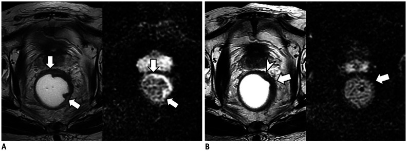
Fig. 5. Cases showing usefulness of DWI for assessment of mrTRG in 80-year-old man with rectal cancer.
A. On T2W axial (left) and DWI (right) before CRT, tumor (arrows) is located between 12 and 5 o'clock position of rectum. Tumor shows intermediate high SI on T2WI (left) and diffusion restriction on DWI (right). B. After CRT, most of tumor (arrow) was replaced by fibrosis with dark SI on T2WI (left). However, there is suspicious area (arrowhead) with intermediate high SI at periphery of tumor. Therefore, two radiologists reported mrTRG as grade 3. However, post-CRT DWI (right) shows completely dark SI at entire tumor (arrow) which suggests absence of viable tumors. Radiologists changed mrTRG into grade 2. On histopathology, there were no tumor cells within fibrosis (not shown). Pathologists finally graded pTRG as grade 2.

