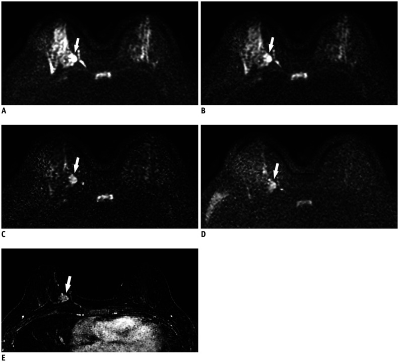
Fig. 1. 49-year-old woman with 2.4 cm invasive ductal carcinoma (histologic grade 2) in right 3 o'clock position.
On conventional axial DWI at b-value of 800 s/mm2 (A) and on synthetic DWI at b-value of 1000 s/mm2 (B), breast cancer (arrows) shows hyperintensity, but border is obscured by insufficiently suppressed normal glandular tissue [suppression of normal glandular tissue: 2, image quality: 3, lesion conspicuity: 2, and cancer-to-parenchyma contrast ratios: 0.33 for (A) and 0.37 for (B)]. On synthetic DWI at b-value of 1500 s/mm2 (C), cancer (arrows) is clearly seen (suppression of normal glandular tissue: 3, image quality: 3, lesion conspicuity: 4, and cancer-to-parenchyma contrast ratio: 0.45) when compared with conventional DWI at b-value of 1500 s/mm2 (D) (suppression of normal glandular tissue: 3, image quality: 3, lesion conspicuity: 3, and cancer-to-parenchyma contrast ratio: 0.39). (E) Axial post-contrast subtraction image at same slice location demonstrates the enhancing tumor (arrow), showing similar lesion visibility on synthetic DWI at b-value of 1500 s/mm2. DWI = diffusion-weighted imaging

