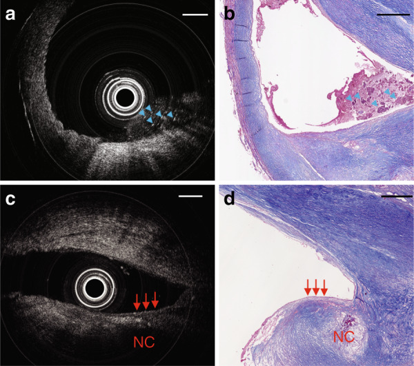
Fig. 4. OCT imaging in a severely diseased human carotid artery.

a Cross-sectional OCT image. b Masson’s trichrome staining of adjacent sections shown in a; blue arrows indicate thrombus that appears to contain fibrin, platelets, and cellular debris. c Another cross-sectional OCT image. d Masson’s trichrome staining of the same area shown in c; red arrows point to a fibrous cap with an adjacent necrotic core. NC: necrotic core. Scale bar: 500 µm
