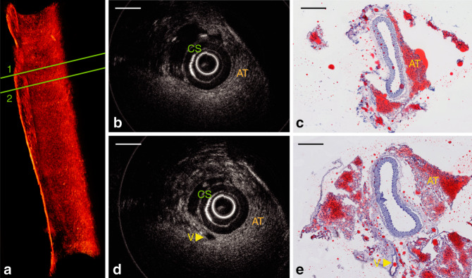
Fig. 5. In situ OCT imaging in a normal mouse aorta.
a Three-dimensional rendering of the volumetric data set acquired with a 3D printed intravascular imaging catheter in a healthy mouse artery with no atherosclerosis. The volume comprises 500 frames of OCT images. A video of this 3D rendering is available as Supplementary Movie 1. b Cross-sectional OCT image of region 1 in a; c corresponding Oil Red O-stained section, where lipid-rich tissue was stained to red color, which indicates the existence of adventitial and perivascular adipose tissue (AT). Tearing in the histology (not OCT) is apparent between the adipose tissue and vessel owing to poor microtome sectioning during histopathology procedures. d Cross-sectional OCT image of region 2 in a; e corresponding Oil Red O-stained sections. AT: adipose tissue that surrounds artery; CS: catheter sheath; V: an additional vessel parallel to the aorta. Scale bar: 250 µm

