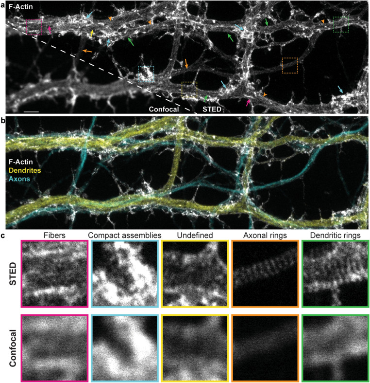
Figure 1.
STED nanoscopy reveals diverse nanostructures of F-actin, in cultured hippocampal neuronal processes, that cannot be resolved with confocal microscopy. (a) Representative image of the F-actin skeleton showing the diversity of nanostructures that can be observed with STED nanoscopy. Arrows point to regions exhibiting dendritic rings (green), axonal rings (orange), longitudinal fibers (magenta), compact assemblies (cyan), or undefined or diffuse signal (yellow). Orange arrowheads indicate regions of overlapping axonal and dendritic patterns. (b) Three color imaging of the region in (a) showing the overlap between axons (cyan, phosphorylated neurofilaments—SMI31) and dendrites (yellow, MAP2). MAP2 and SMI31 were imaged with confocal resolution to highlight the shape of the processes. (c) Insets show a magnification of the regions indicated with the dashed squares in (a) for both STED (top) and confocal (bottom) imaging modalities. Scale bar 2 m, insets .

