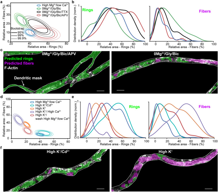
Figure 7.
Synaptic NMDAR activity and influx drive a reversible dendritic F-actin reorganization from a ring to fiber pattern. (a) Mean distributions of dendritic F-actin rings and fibers using bootstrapping for synaptic stimulation (0/Gly/Bic for 10 min) without (green) or with TTX (black) or APV (red) compared with the low activity high /low condition (blue). (b) Density distribution of the raw data. TTX (1 M) partially but significantly blocks the F-actin remodeling caused by 0/Gly/Bic stimulation, while APV (25 M) blocks it further ( and , respectively). (c) Representative images of neurons treated with 0/Gly/Bic with (left) and without (right) APV segmented with our deep learning based approach. (d) Mean distributions of dendritic F-actin rings and fibers using bootstrapping for 2 min high stimulation (1.2 mM , orange) or with 2.4 mM (violet) or with 1.2 mM and 50 M (cyan). The red circles indicate the same high stimulation (1.2 mM ) condition followed by 15 min wash in high /low . The F-actin remodeling is -dependent and reversible, at least partially within 15 min. (e) Density distribution of the raw data. (f) Representative images of neurons treated with high stimuli with (left) and without (right) segmented with our deep learning based approach. Statistical analysis performed with a randomization test (see Materials and Methods). Number of independent cultures (N) and number of neurons (n): high /low , , 0/Gly/Bic , ; 0/Gly/Bic M TTX , ; 0/Gly/Bic + M APV , ; high /low ; high mM , ; high mM , ; high M , ; high min wash high , . For the raw images without overlay see Supplementary Fig. 14.

