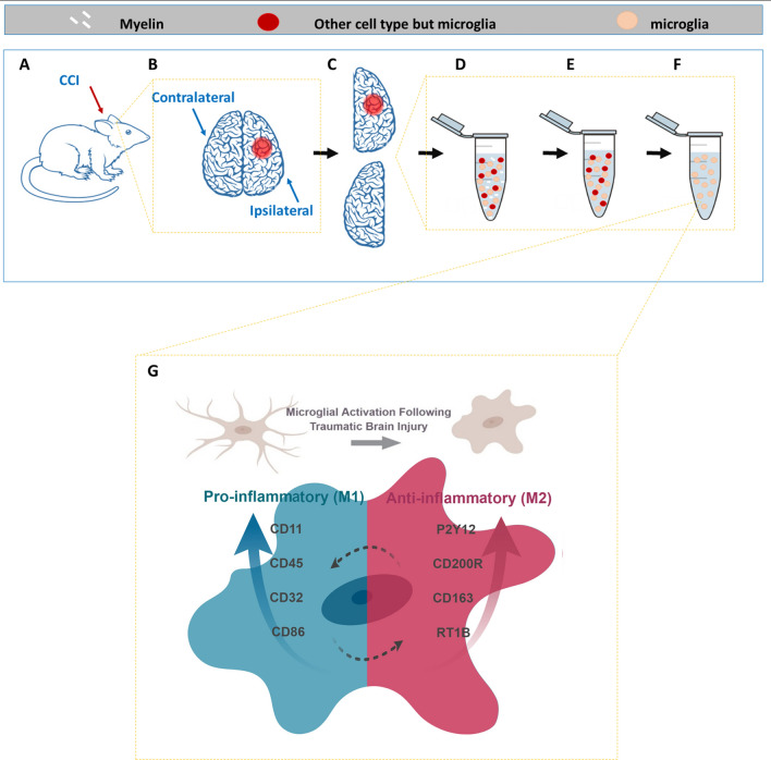
Figure 1.
Process and Rationale Illustration. (A) Rats were injured using the controlled cortical impact method, to create a precise injury at one hemisphere. (B) Each hemisphere was processed to a separate sample, by mechanically and enzymatically digesting the tissue into single cell suspension (D), myelin removal (E) and microglial (CD11bc) enrichment (F), which is the final sample that was later analyzed with flow cytometry. (G) Considering the morphological changes and the changes in markers expression we used a validated multi-parametric flow cytometry panel to evaluate all parameters together to create a profile of the activated microglia at 24 after injury. Made in ©BioRender—biorender.com.

