Figure 1.
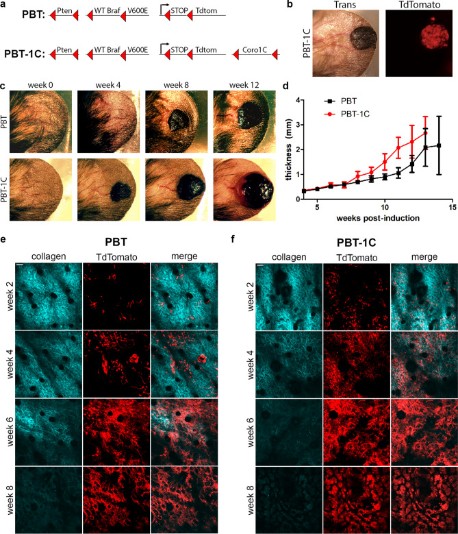
Coronin 1C-null melanoma grows faster than endogenous-expressing tumors. (a) Schematic diagram of two GEM models, PBT and PBT-1C. Upon 4-hydroxytamoxifen application, CreER expressed under the melanocyte-specific Tyrosinase promotor excises the DNA between LoxP sites, denoted with red triangles, resulting in Pten deletion, BRAF constitutive activation and TdTomato expression. In the PBT-1C model, Coro1C is also deleted. (b) Transillumination and red fluorescence for TdTomato imaging of a PBT-1C primary tumor 8 weeks after induction with 4-hydroxytamoxifen. (c) Transillumination of one PBT primary tumor and one PBT-1C primary tumor over a 12-week period following induction. (d) Quantification of the mean thickness + /− 95% CI of the ear and primary tumor (N of 15 for PBT and N of 16 for PBT-1C. (e, f) Representative multi-photon imaging of second harmonic signal of the bundled collagen layer and TdTomato expression in recombined melanocytes in the primary tumor of a PBT mouse ear (e) and a PBT-1C mouse ear (f) over an 8-week period post induction with 4-HT. Scale bars = 50 µm.

