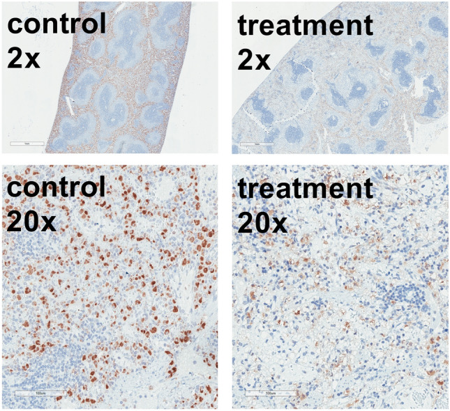
Figure 4.

Macrophage staining of the spleen. Immunohistochemical staining with CD68 clone ED1 antibody of spleen tissue of animals diluted using 5% HSA (control) or 12 vol% capsules (treatment). Hematoxylin (blue) co-staining was used for orientation. The brown colour represents the macrophages. Control animals showed unaffected macrophages in contrast to foamy vacuolized macrophages in the treatment group. Staining was performed with n = 8 spleens in each group. Treatment spleens demonstrated lost tissue structure within the red pulpa with foamy, swollen macrophages. The white pulpa was not affected. The control spleens were intact.
