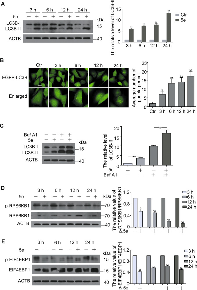
Fig. 3. Compound 5e induced autophagy in a mTOR-dependent manner.
a Western blot analysis of LC3B-I and LC3B-II in A549 cells treated with compound 5e at 10 μM for indicated times and quantification of LC3B-II levels. b Images of EGFP-LC3B U87 cells were treated with compound 5e at the concentration of 10 μM for 3, 6, 12, and 24 h (200×) and quantification of EGFP-LC3B dots. Bar = 10 μm. c Western blot analysis of LC3B-I and LC3B-II in A549 cells treated with compound 5e (10 μM), Baf-A1 (50 nM), or both for 12 h and quantification of LC3B-II levels. d Western blot analysis of RPS6KB1 and p-RPS6KB1 (S424/T421) in A549 cells treated with compound 5e at 10 μM for indicated times and quantification. e Western blot analysis of EIF4EBP1 and p-EIF4EBP1 (S65/T70) in A549 cells treated with 5e at 10 μM for indicated times and quantification. β-actin was used as a loading control. Results were presented as mean ± SE; n = 3; *p < 0.05; **p < 0.01.

