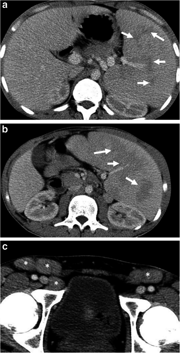
Fig. 16.

A 21-old man was admitted to the emergency department with a 2-week history of left upper quadrant abdominal pain and fever. Physical examination revealed splenomegaly and inguinal lymphadenopathy. Increased serum levels of acute-phase reactants, leukocytosis, and high sedimentation were evident at blood analysis. a, c Axial contrast-enhanced portal venous phase CT images demonstrate patchy areas of hypoattenuation (arrows, a, b) within the enlarged spleen and bilateral inguinal lymphadenopathy (asterisks, c). The liver was normal except for mild hepatomegaly (18 cm). The diagnosis of Leishmaniasis was made by histopathological examination following splenectomy
