
Fig. 6.
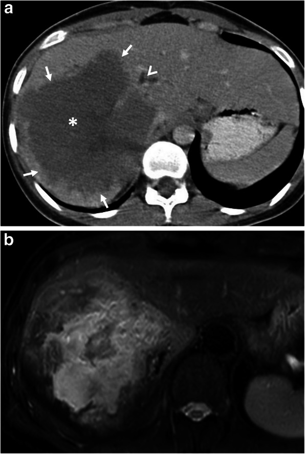
A 21-year-old woman was admitted to the emergency department with a 2-week history of jaundice and upper right quadrant pain. a Axial contrast-enhanced CT image demonstrates a heterogeneous infiltrative liver mass (asterisk) with irregular margins (arrows). The presence of biliary dilatation (arrowhead) due to compression of the mass was also noted. The diagnosis of alveolar echinococcosis was made by histopathological examination. b Fat saturated T2-weighted MR-image demonstrates internal heterogeneity of the mass mimicking primary liver malignancy. Chest CT findings were unremarkable for hydatid disease (not shown)