Fig. 8.

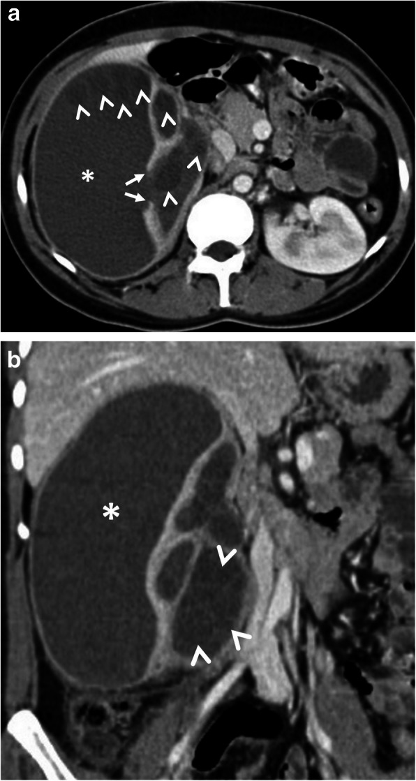
A 24-year-old woman was admitted to the emergency department with an acute onset of right flank pain and fever. Increased serum levels of acute-phase reactants, leukocytosis, and elevated serum creatinine levels were evident at blood analysis. a, b Axial (a) and coronal (b) contrast-enhanced CT images demonstrate a large renal subcapsular Gharbi type 3 hydatid cyst (asterisks) rupture into the pelvicalyceal system. Loss of integrity of the renal parenchyma found on CT image indicates the site of rupture (arrows, a). Daughter cysts within the cyst and also in the pelvicalyceal system are noted (arrowheads, a, b). The diagnosis was confirmed by histopathological examination following surgery
