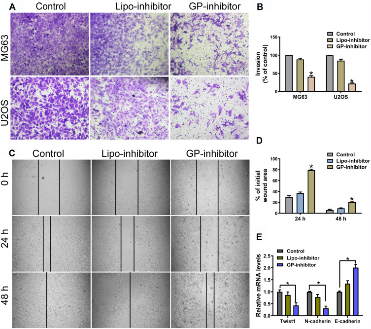
Figure 5.
GP inhibitor inhibits invasion and migration in MG63 and U2OS cells. (A) The invasion of MG63 and U2OS cells were measured for 24 h using a Matrigel-coated chamber with polycarbonate filters. (B) Statistical analysis of the invasion cell ratio is presented for each group. *p < 0.05. (C) The wounds of confluent MG63 cells were created with 100 μL yellow tips and incubated with a lipo-inhibitor or GP-inhibitor (5 μg/mL). Images of the wound at different time points are shown, and the borders of the wound are shown by a pair of black lines. (D) Statistical analysis of cell migration by calculating the ratio of the final cell-free gap to the initial wound area. *p < 0.05. (E) The expression levels of twist1, N-cadherin and E-cadherin after treatment with lipo-inhibitor or GP-inhibitor were evaluated by qRT-PCR. *p < 0.05.

