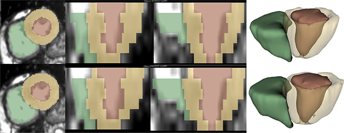
FIG. 1.
Segmentation results for LV blood-pool, LV myocardium, and RV blood-pool. First column shows the short-axis view, second and third columns show orthogonal long-axis views, and the fourth column shows generated three-dimensional models. Reference (top row) and segmentation obtained from the DMR-UNet model (bottom row). [Color figure can be viewed at wileyonlinelibrary.com]

