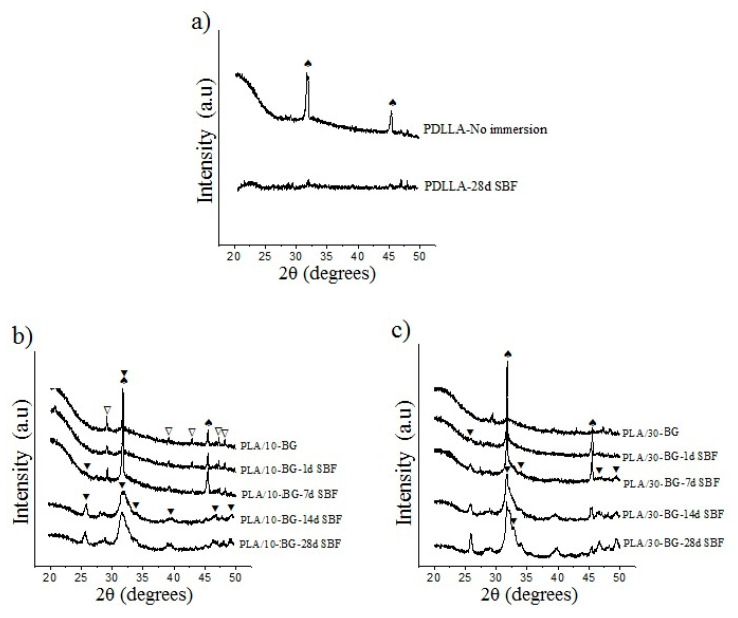
Figure 3.
X-ray diffractograms showing apatite formation on the surface of the scaffolds with bioactive glasses (BG) after immersion in simulated body fluid (SBF). Scaffolds (a) neat PDLLA (b) PLA/10-BG and (c) PLA/30-BG. Crystal phases are labeled as: (⯆) apatite, (∇) CaCO3 (JCPDS 01-085-1108) and (♠) Halite (NaCl, JCPDS 5-0628).

