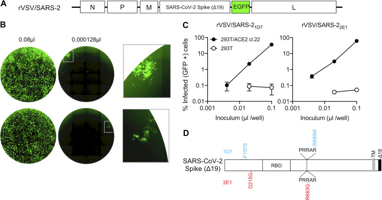
Figure 3.
A replication-competent VSV/SARS-CoV-2 chimera. (A) Schematic representation of the rVSV/SARS-CoV-2/GFP genome in which G-encoding sequences were replaced by SARS-CoV-2 SΔ18 coding sequences. GFP-encoding sequences were introduced between the SARS-CoV-2 SΔ18 and L open reading frames. (B) Representative images of 293T/ACE2(B) cells infected with the indicated volumes of plaque-purified, adapted derivatives (2E1 and 1D7) of VSV/SARS-CoV-2/GFP following passage in the same cell line. Left and center images show contents of an entire well of a 96-well plate, and the right image shows an expanded view of the boxed areas containing individual plaques. (C) Infectivity measurements of rVSV/SARS-CoV-2/GFP virus stocks on 293T/ACE2(B) or control 293T cells, quantified by measuring the percentage of GFP-positive cells at 16 h after infection. Mean and range from two technical replicates are shown. (D) Schematic representation of the adaptive changes acquired in rVSV/SARS-CoV-2/GFP during passage. Changes in 1D7 and 2E1 are shown in blue and red, respectively. PRRAR, amino acid sequence at the furin cleavage site; TM, transmembrane domain.

