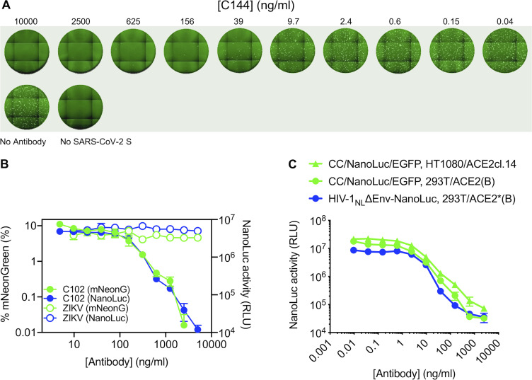
Figure S4.
Examples of neutralization of HIV-1 and VSV pseudotyped virus particles by mAbs targeting SARS-CoV-2 S. (A) Images of Huh7.5 cells following infection with rVSVΔG/NG-NanoLuc pseudotyped virus (∼103 IU/well) in the presence of the indicated concentrations of a human mAb (C144) targeting SARS-CoV-2 S RBD. Images of the entire well of a 96-well plate are shown. (B) Quantification of rVSVΔG/NG-NanoLuc pseudotyped virus infection (measured by flow cytometry (percentage of mNeonGreen-positive cells, green) or by NanoLuc luciferase activity (RLU, blue) in the presence of the indicated concentrations of a human mAb (C102) targeting SARS-CoV-2 S RBD or a control mAb against the Zika virus envelope glycoprotein. Mean and range of two technical replicates are plotted. (C) Quantification of HIV-1NLΔEnv-NanoLuc or CCNanoLuc/GFP pseudotyped virus infection on the indicated cell lines in the presence of the indicated concentrations of a human mAb (C121) targeting SARS-CoV-2 S RBD infectivity was quantified by measuring NanoLuc luciferase levels (RLU). Mean and range of two technical replicates are plotted.

