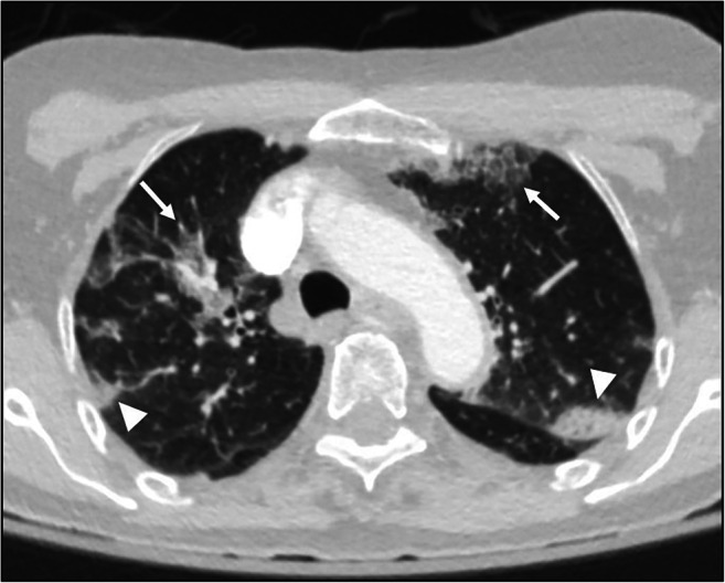
Fig. 2.

Axial image from a non-contrast cervical spine CT in an 84-year-old female presenting after an unwitnessed fall at home and unknown COVID-19 status. Images of the lung apices demonstrate peripheral, multifocal ground glass opacities (white arrows) with areas of consolidation (white arrowheads), a “typical appearance” of COVID-19 [2]. The patient subsequently tested COVID-19 positive
