
Figure 6.
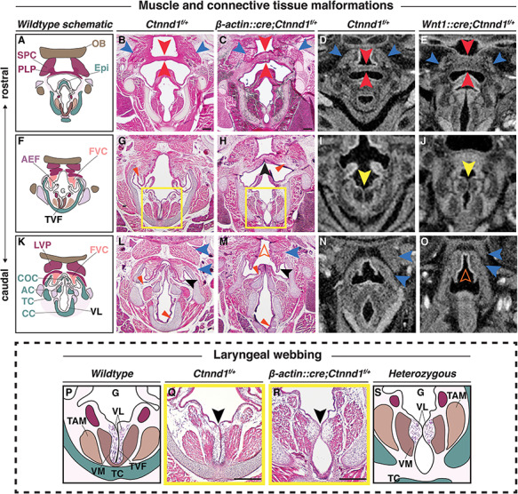
Heterozygous loss of p120-catenin leads to structural changes in the laryngeal apparatus. (A–O) Progression of the pharyngeal and laryngeal anomalies, (A, F and K) Schematics show the organization of the wild-type oropharynx from the more rostral (A) to caudal (K) planes. H&E staining of coronal sections through control (B, G, L: Ctnnd1fl/+) and heterozygous mutants (C, H, M: β-actin::cre/+; Ctnnd1fl/+) littermate at postnatal stage (P1). (B and C) The SPC (blue arrowhead) and PLP (red arrowhead) in mutants are disorganized with an increased thickness in the PLP cranio-caudally (C) as compared with the controls (B). (G and H) The FVC (vestibular folds) are well defined in the controls with abundant ligaments (G, red arrowhead). The FVC are fused in the mutant mice (H, black arrowhead) with ill-defined vestibular ligaments (H, red arrowhead). (L and M) The muscle attachments (blue arrowheads) superior to the FVC (black arrowhead) are well organized bilaterally in the controls surrounding the COC (L). Caudally, when the FVC separated in the mutants, it appeared hypoplastic (black arrowhead) as did the COC. The muscles (blue arrowheads) were ectopically fused to the LVP, producing an appearance of a ‘high-arched’ epiglottal area (M, orange hollow arrowhead). (D, E, I, J, N and O) Neural crest-specific mutants showed comparable laryngeal phenotype. μCT soft tissue scans of E16.5 control (D, I, N: Ctnnd1fl/+) or neural-crest-specific (E, J, O: Wnt1::cre/+; Ctnnd1fl/+) heterozygous mutant littermates. (D and E) Compare the PLP in control (D) to the very thick PLP muscle seen in mutant (E, red arrowheads). Compare the SPC in control (D) to the disorganized and hypoplastic SPC muscles seen in mutants (E, blue arrowheads). (I and J) Laryngeal webbing was observed in mutant TVF (J, yellow arrowhead) compared with parallel TVF in control littermate (I, yellow arrowhead). (N and O) Note aberrant muscle attachments (blue arrowheads) in (O) compared with control (N). Control (N) epiglottal region compared with the high-arched epiglottal area observed in mutant littermate (O, orange hollow arrowhead). (P–S) The laryngeal webbing phenotype. (P and S) Schematic representations of the wild-type (P) and mutant (S) anatomy at the vocal folds (TVF) from yellow-boxed insets in (G) and (H), respectively. (Q and R) H&E staining of coronal sections through control (Q: Ctnnd1fl/+) and heterozygous mutant (R: β-actin::cre/+;Ctnnd1fl/+) littermate at P1. (Q) In controls, well-defined VLs run parallel to the true vocal fold/cords (TVF). Underlying, the vocalis muscle (VM) and the thyroarytenoid muscle (TAM) are clearly attached and well-organised. (R) Laryngeal webbing is seen in the heterozygous mutant mice, where the VLs accumulate at a thin contact point (black arrowhead), thus perturbing the correct muscle attachments of the VM and TAM. Scale bars = 100 μm. Abbreviations: PLP, palatopharyngeus muscle; TAM, thyroarytenoid muscle; VM, vocalis muscle; HB, hyoid bone; Epi, epiglottis; OB, occipital bone; LVP, levator veli palatini muscle; AEF, aryepiglottic fold; FVC, false vocal cord; CC, cricoid cartilage; TC, thyroid cartilage; AC, arytenoid cartilage.