Figure 7.

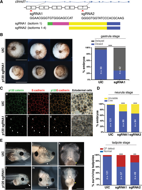
Ctnnd1 knockouts in Xenopus give rise to craniofacial and heart defects. (A) Embryos were injected at the one-cell stage with sgRNAs, sgRNA1 and sgRNA2 targeting exons 3 and 7, respectively. (B) Ventral view showing blastopores at stage 11. Embryos injected with sgRNA1 had delayed blastopore closure (bottom row) compared to un-injected controls (UIC) (top row). The bar chart shows quantitation. Scale bars = 100 μm. (C) Confocal sections through the apical surface of ectodermal cells at stage 11 of embryos injected with sgRNA1 (e–h) and UICs (a–d). (C) (a–d) p120-catenin (a, green) is expressed in puncta at the cell membranes. E-cadherin (b, red) is expressed more evenly through the cell membranes. Both are colocalized at the cell–cell interface (c, d). Endogenous levels of p120-catenin and E-cadherin are diminished at the cell–cell interface in the sgRNA1-injected embryos (e, f). Residual p120-catenin and E-cadherin are seen in a spot-like pattern, only at the tricellular junctions (e–h, white arrowheads). (D) p120-catenin depletion led to lethality in embryos by the neurula stage. (E) Stage 46 tadpoles. (E) (i, l) Lateral views show a flattened profile in p120 CRISPR tadpoles (l) compared with UICs (i). (E) (j, m) Frontal views showing a reduction in the size of mouth opening and a persistent cement gland (white arrowhead) in p120 CRISPR tadpoles (m) compared with UICs (j). (E) (k, n) Ventral views showing a reduction in the size of craniofacial cartilages, altered cardiac looping (black-dashed outline) and altered gut coiling (yellow arrowhead) in p120 CRISPR tadpoles (n) compared to UICs (k). Quantification of craniofacial defects in UIC and p120 depleted tadpoles. Scale bars = 100 μm. ****P < 0.0001; ***P < 0.001.
