Figure 3.
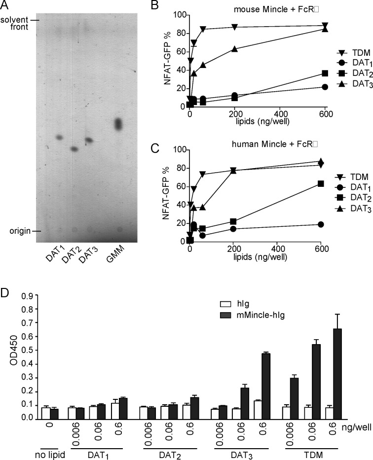
Synthetic DAT3 is recognized by human and mouse Mincle. (A) Before functional assays, DAT1, DAT2, and DAT3 were analyzed by thin-layer chromatography for relative quantification and the presence of major breakdown products. (B and C) NFAT-GFP reporter cells expressing mouse Mincle + FcRγ or human Mincle + FcRγ were stimulated with the indicated amount of DAT1, DAT2, DAT3, or TDM. After 24 h, induction of NFAT-GFP was analyzed by flow cytometry. (D) ELISA-based detection of DAT1, DAT2, DAT3, or TDM by mouse Mincle-human Ig Fc (mMincle-hIg) fusion proteins. Bound protein was detected with antihuman Ig-horse radish peroxidase (HRP), followed by the addition of a colorimetric substrate and measurement.

